2024 | Volume 25 | Issue 2
Author: Professor Richard C Turner

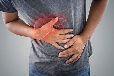
Severe acute pancreatitis can be fatal, due to a storm of inflammatory mediators often with underlying comorbidities. Over the three-year period from 2021 to 2023, the Australian and New Zealand Audit of Surgical Mortality (ANZASM) undertook 162 First Line Assessments for pancreatitis deaths, of which 14 led to Second Line Assessments because areas of concern related to pre-or in-hospital management. Such cases represent the ‘tip of the iceberg’ of a disease process that is largely self-limiting and only requires surgery, in the form of cholecystectomy, if gallstones are the precipitating cause. Nevertheless, the overall disease burden of acute pancreatitis on the health system cannot be discounted. In a typical Acute Surgical Unit, such as at the Royal Hobart Hospital, acute pancreatitis cases occupy up to 25 per cent of bed capacity at any one time. Although the global incidence of acute pancreatitis is increasing, little data exists about trends in Australia.
A recent population-based study has for the first time objectively quantified the disease burden of pancreatitis in an Australian state jurisdiction, Tasmania. The Tasmanian Data Linkage Unit derived the study cohort by probabilistically linking individuals across seven public hospital and pathology laboratory datasets. In a 12-year period from 2007 to 2018, 4905 episodes of acute pancreatitis were identified, of which 3503 (71.4 per cent) were first episodes. There was an approximately equal distribution of males and females, with a mean age at first presentation of 55 years. In terms of aetiology, 58 per cent of first episodes where coded as unspecified, 26 per cent as biliary, and 13 per cent as alcohol-related. The remaining three per cent were a combination of other, drug-induced, and idiopathic aetiologies. First episodes had a median four-day length of hospital stay; four per cent required ICU admission, although only two per cent died.
At 54.2 per 100,000 people per year, the incidence of acute pancreatitis in Tasmania over the 12-year study period was comparable to the 58.4 per 100,000 people per year in a 2017 study from Aotearoa New Zealand[1]. It was also well above the combined value of 33.7 per 100,000 people per year from a 2016 meta-analysis of a series of 10 population-based cohort studies worldwide[2]. The assessed Tasmanian incidence rate may yet be an underestimate given the unavailability of private hospital data. The findings also concur with Australian[3] and global reports[4],[5] that the annual incidence of acute pancreatitis has been consistently increasing over the last few decades, presumably due to the growing worldwide prevalence of known and unknown aetiological factors[6].
As with the studies from Wales[7] and Aotearoa New Zealand1,the Tasmanian study also revealed differential incidence rates across socio-demographic groups. In particular, there appears to be an inverse gradient between incidence and socio-economic status, based on the Australian Bureau of Statistics’ (ABS) Index of Relative Socioeconomic Disadvantage (IRSD). This is a composite of economic and social conditions within an area, encompassing features such as unemployment, education, housing, and disability. A high IRSD score indicates a relative lack of disadvantage.
The annual incidence rates for the lowest three IRSD quartiles also showed a consistent linear increase over the 12-year study period, while the highest (least disadvantaged) quartile actually showed a slight overall decline in incidence. These results suggest that those in the least disadvantaged quartile are decreasingly exposed to the factors that continue to drive the increasing incidence in the more disadvantaged groups.
The relationship between acute pancreatitis and socio-economic status is complex and likely mediated by the prevalence of aetiological factors in the population residing in a particular area. Dietary patterns characterised by increased energy intake with highly refined sugars and sweet foods, high fructose intake, low fibre, high fat, fast food consumption, and low vitamin C intake increase the risk of gallstone (and sludge or microcrystal) formation[8],[9] as do certain genetic and epigenetic factors[10]. For alcohol-related pancreatitis, individual susceptibility may be increased by factors such as smoking, obesity, and various genetic mutations[11]. According to the ABS’ National Health Survey 2017-2018[12], the prevalence of behavioural or lifestyle risk factors varies with IRSD score, although not always in the same direction. While smoking, overweight/obesity, inadequate fruit and vegetable intake, and consumption of sweetened drinks are more prevalent in the lowest IRSD quintiles, the reverse is true for alcohol consumption exceeding guideline recommendations.
The study is unable to conclude that the common aetiologies, namely gallstones and alcohol, are not driving the temporal increase in incidence, which is largely attributable to acute pancreatitis of ‘unspecified’ aetiology. International consensus guidelines recommend that, wherever possible, an aetiological attribution should be sought. However, it is often not possible for clinicians to commit to a specific aetiology.
Certain aetiological categories with relatively subjective diagnostic criteria may be coded as ‘unspecified’ if the discharging clinician has any uncertainty. This is especially the case for alcohol-related pancreatitis. Despite consensus that an acute episode only occurs after at least five years of four to five standard drinks per day[13], the threshold may be lower for some people. Further, self-reported alcohol intake is often underestimated[14]. In addition, biliary sludge and microlithiasis, known causes of biliary AP[15],[16], are poorly detected by transabdominal ultrasound[17]. Endoscopic ultrasound (EUS) is considered the gold standard for diagnosis of biliary sludge[18]; however, it is more invasive and expensive and, if available, is mostly reserved for recurrent episodes. Moreover, it is acknowledged that both acute and chronic pancreatitis may be a result of multiple risk factors acting synergistically[19]. Finally, it should be noted that 29 per cent of unspecified cases were only identified in the ED dataset, where ICD-10 coding did not ascribe aetiological sub-categories and coding may not have been as rigorous as for Admitted Patient data[20]. Many of these unspecified cases from ED may have been cases of biliary AP that were subsequently admitted to a private hospital for cholecystectomy, although this is a matter of speculation.
In summary, the incidence of acute pancreatitis in the Tasmanian population is high and continues to increase. We found a complex and significant correlation between incidence rates and socio-economic status, likely driven by the prevalence of known and unknown risk factors. To better define these risk factors, greater efforts should be given to attributing aetiology for all new admissions.
References
[1] Pendharkar SA, Mathew J, Zhao J, Windsor JA, Exeter DJ, Petrov MS. Ethnic and geographic variations in the incidence of pancreatitis and post-pancreatitis diabetes mellitus in New Zealand: a nationwide population-based study. N Z Med J. 2017;130(1450):55 - 68.
[2] Xiao AY, Tan MLY, Wu LM, Asrani VM, Windsor JA, Yadav D, et al. Global incidence and mortality of pancreatic diseases: a systematic review, meta-analysis, and meta-regression of population-based cohort studies. Lancet Gastroenterol Hepatol. 2016;1:45 - 55.
[3] Ah-Tye PJ. Pancreatitis in remote Australia: an indigenous perspective. Australian Journal of Rural Health. 2001;9(3):134 - 7.
[4] Iannuzzi JP, King JA, Leong JH, Quan J, Windsor JW, Tanyingoh D, et al. Global incidence of acute pancreatitis is increasing over time: A systematic review and meta-analysis. Gastroenterology. 2022;162:122 - 34.
[5] Han K, Chen S, Song Y, Du C, Gao F, Liu S, et al. Burden of pancreatitis and associated risk factors in China, 1990 to 2019: a systematic analysis for the Global Burden of Disease Study 2019. Chinese Medical Journal. 2022;135(11):1340 - 7.
[6] Barreto SG, Habtezion A, Gukovskaya A, Lugea A, Jeon C, Yadav D, et al. Critical thresholds: key to unlocking the door to the prevention and specific treatments for acute pancreatitis. Gut. 2021;70:194 - 203.
[7] Roberts SE, Akbari A, Thorne K, Atkinson M, Evans PA. The incidence of acute pancreatitis: impact of social deprivation, alcohol consumption, seasonal and demographic factors. Alimentary Pharmacology and Therapeutics. 2013;38:539 – 48.
[8] Di Ciaula A, Garruti G, Frühbeck G, De Angelis M, de Barie O, Wang DQ-H, et al. The role of diet in the pathogenesis of cholesterol gallstones. Curr Med Chem. 2019;26(19):3620 – 38.
[9] Jessri M, Rashidkhani B. Dietary patterns and risk of gallbladder disease: A hospital-based case-control study in adult women. J Health Popul Nutr. 2015;33(1):39 - 49.
[10] Di Ciaula A, Wang DQ-H, Portincasa P. An update on the pathogenesis of cholesterol gallstone disease. Curr Opin Gastroenterol. 2018;34(2):71 - 80.
[11] Wilson JS, Pirola RC, Apte MV. Epidemiology and Etiology of Alcohol-Induced Pancreatitis. In: Beger HG, Buchler MW, Hruban RH, Mayerle J, Neoptolemos JP, editors. The Pancreas. 4th ed: Wiley Blackwell; 2023.
[12] Australian Bureau of Statistics. National Health Survey: First Results, 2017–2018. In: Australian Government 2018, editor.
[13] Klochkov A, Kudaravalli P, Lim Y, Sun Y. Alcoholic Pancreatitis. Treasure Island (FL): StatPearls Publishing; 2023 16 May 2023.
[14] Hesselbrock M, Babor TF, Hesselbrock V, Meyer RE, Workman K. “Never Believe an Alcoholic”? On the Validity of Self-Report Measures of Alcohol Dependence and Related Constructs. The International Journal of the Addictions. 1983;18(5):593 - 609.
[15] Lee SP, Nicholls JF, Park HZ. Biliary sludge as a cause of acute pancreatitis. N Engl J Med 1992;326(9):589 - 93.
[16] Ros E, Navarro S, Bru C, Garcia-Pugés A, Valderrama R. Occult microlithiasis in “idiopathic” acute pancreatitis: prevention of relapses by cholecystectomy or ursodeoxycholic acid therapy. Gastroenterology. 1991;101:1701 - 9.
[17] Ko CW, Sekijima JH, Lee SP. Biliary sludge. Ann Intern Med. 1999;130:301 - 11.
[18] Żorniak M, Sirtl S, Beyer G, Mahajan UM, Bretthauer K, Schirra J, et al. Consensus definition of sludge and microlithiasis as a possible cause of pancreatitis. Gut 2023;0:1–8 doi:101136/gutjnl-2022-327955.
[19] Weiss FU, Laemmerhirt F, Lerch MM. Etiology and Risk Factors of Acute and Chronic Pancreatitis. Visc Med. 2019;35:73 - 81.
[20] Tran QN, Lambeth LG, Sanderson K, de Graaff B, Breslin M, Huckerby EJ, et al. Trend of emergency department presentations with a mental health diagnosis in Australia by diagnostic group, 2004–05 to 2016–17. Emergency Medicine Australasia. 2020;32:190 - 201.